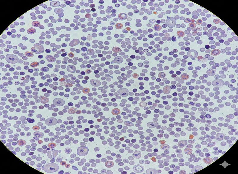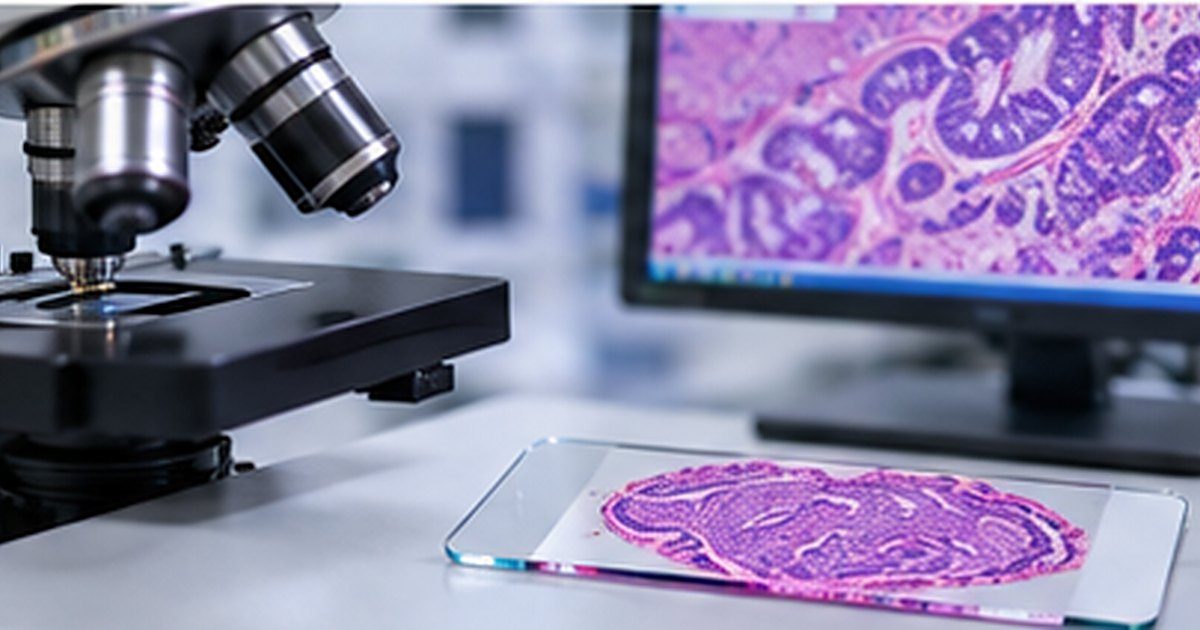
Digital pathology offers an important shift in how histopathology services store, review and share tissue images. Whole-slide scanners turn glass slides into high-resolution files that can be examined on validated digital workstations. The potential benefits include easier remote collaboration, access to archived material and integration with image analysis tools.
But buying a scanner does not, by itself, create a safe digital pathology service. The transition changes work across histology preparation, scanning, image quality assurance, pathologist reporting, information technology and data governance.
The Royal College of Pathologists and UK screening guidance emphasise validation, training, ongoing quality assurance and suitable technical infrastructure. Successful adoption depends on treating the change as a laboratory-wide clinical programme.
At a glance: Digital pathology implementation involves more than scanning glass slides. A safe clinical service needs fit-for-purpose scanners, image quality checks, secure storage, validated workflows, trained reporting staff, interoperability and contingency arrangements. The intended clinical use should guide the validation plan and operational design.
Begin with a defined clinical use
The first question is what the digital system will be used for. A department may want to support multidisciplinary meetings, education, second opinions, remote reporting or primary diagnosis. Each has different risks and requirements.
A system used for teaching images has different implications from one used for issuing patient diagnoses. The laboratory should agree the initial scope, any excluded specimen types or techniques, and the conditions under which glass-slide review remains necessary.
It is sensible to distinguish whole-slide imaging from algorithmic analysis. A scanner and viewer may support a digital workflow without any artificial intelligence. If image analysis software is added, its intended purpose, regulatory status, local evaluation and governance need separate consideration.
Follow the slide through the entire process
Before an image reaches a reporting screen, the specimen has been processed, embedded, sectioned, stained, coverslipped, labelled and scanned. Poor section quality, staining artefacts, barcode problems or scanner failures can undermine the result before a pathologist opens the file.
The project should define how slides enter the scanner queue, how images are linked to the correct case, who checks scan quality and what happens when rescanning is required. Pathways for urgent material, special stains and immunohistochemistry should be considered explicitly rather than assumed to work like routine haematoxylin and eosin slides.
Storage also matters. Whole-slide images can be large, and institutions must consider retention obligations, access, backup, network performance and the lifecycle cost of image archives. The clinical reporting workflow should not be dependent on a fragile manual file-transfer process.
Validate the people and the system
Digital diagnosis requires an appropriate programme of equipment acceptance, technical verification and pathologist training and validation. In practical terms, each reporting pathologist needs to become familiar with the digital system and demonstrate that they can use it safely for the relevant scope of work.
Cases for validation should represent the actual specimens and diagnostic tasks the pathologist will encounter. Discrepancies between digital and glass review require investigation and documentation. Performance should be revisited when material changes are made to viewing equipment, image formats, software or the intended clinical workflow.
England's 2025 guidance on digital histopathology in cancer screening points providers towards the Royal College of Pathologists' implementation recommendations and stresses maintenance and optimisation of scanners and monitors. It also places responsibility for training and validation on the provider.
Design a resilient reporting environment
Workstation ergonomics, monitor characteristics and image loading speed all affect reporting. A technically excellent image is of limited use if navigation is slow, access is unreliable or the system makes it difficult to compare slides in a case.
Cybersecurity, patient confidentiality and access controls need consideration from the outset. Departments should test what happens when the network or image archive is unavailable. Where digital reporting becomes essential to service delivery, a documented fallback is a patient-safety measure.
Digital platforms can also create new opportunities for collaboration across sites. However, sharing a slide does not replace the need for clear reporting responsibility, clinical governance and data-sharing agreements.
Evaluate benefit after deployment
Digital pathology should be judged against agreed objectives rather than the number of scanned slides. Potential measures include rescan rates, image quality failures, report availability, pathologist experience, retrieval time, referral workflows and operational resilience.
Some benefits may take time to emerge, and some departments may retain a hybrid workflow. The goal should be safe, clinically useful adoption, not digital conversion as an end in itself.
Where Optymum SS fits: Optymum SS can support digital pathology readiness assessments, workflow mapping, technology evaluation and implementation documentation. Clinical diagnostic validation and reporting remain the responsibility of the relevant pathology service and authorised professionals.
Related on this site: Cellular Pathology or Histopathology, Smart Lab & Digital Solutions.
Key takeaways
- Start with intended clinical use and governance.
- Validate whole-slide workflows, not just image capture.
- Monitor quality, interoperability and service resilience after go-live.
Frequently asked questions
What is digital pathology?
Digital pathology uses digitised pathology slides and related systems for review, sharing, analysis and, where validated and authorised, clinical reporting.
Can a laboratory start digital reporting after buying a scanner?
Not automatically. The laboratory must assess and validate the full intended workflow, including image quality, display equipment, software, staff, information governance and fallback processes.
How should digital pathology benefits be assessed?
Measure the actual workflow: scanning failures, reporting turnaround, retrieval time, consultation pathways, quality issues and total operating costs.